
Electrophoresis
Electrophoresis chambers are essential tools used in molecular biology for separating DNA, RNA, and proteins based on their size and charge. The principle behind electrophoresis is the migration of charged particles (such as DNA or proteins) in an electric field, with molecules moving towards the electrode with an opposite charge. Here’s a breakdown of the key components and considerations when it comes to electrophoresis chambers for DNA and protein gels:
key components of Electrophoresis :
DNA Electrophoresis Chambers :
Purpose : Primarily used to separate DNA fragments by size, based on their ability to move through a gel matrix (usually agarose).
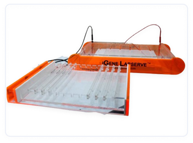
Materials : Typically made from clear, non-conductive plastic (e.g., acrylic) for visibility.
Key Components :
Gel Tray : Where the agarose gel is cast.
Buffer Solution : Provides an environment that maintains the conductivity of the gel. Common buffers include TAE (Tris-acetate-EDTA) and TBE (Tris-borate-EDTA).
Electrodes : Provide the electric field to drive DNA migration. Anode (+) and cathode (−) are placed at either end of the gel chamber.
Power Supply : Provides the electric current necessary to drive electrophoresis. Voltages vary depending on the gel size, typically ranging from 70 to 150 V.
Loading Wells : Where DNA samples are loaded into the gel.
Gel : The medium, usually agarose, that supports the DNA fragments during migration.
Applications :
PCR Product Analysis : To check the size of PCR products.
Restriction Digestion : To separate DNA fragments produced by restriction enzymes.
DNA Ladder : To determine the size of the unknown DNA fragments by comparison to a standard marker.
Protein Electrophoresis Chambers :
Purpose : Used to separate proteins based on their size (molecular weight) and charge. This is often done using SDS-PAGE (Sodium Dodecyl Sulfate PolyAcrylamide Gel Electrophoresis) for denaturing protein analysis.
Materials : Similar to DNA electrophoresis chambers, protein electrophoresis chambers are typically made from transparent materials for clear visualization.
Key Components :
Polyacrylamide Gel : Used as the separation matrix for proteins. The gel is usually made by polymerizing acrylamide and bisacrylamide in the presence of a crosslinker.
Buffer System : Common buffer systems include running buffers such as Tris-Glycine-SDS (for SDS-PAGE).
Electrodes : Like DNA electrophoresis, electrodes apply the electric field.
Loading Wells : Where protein samples are loaded.
Power Supply : Provides the necessary current, typically in the range of 100–150 V for SDS-PAGE.
Applications :
Protein Size Determination : SDS-PAGE allows determination of protein size by comparing to a molecular weight marker.
Western Blotting : After separation via SDS-PAGE, proteins are transferred to a membrane for further analysis (e.g., antibody probing).
Piezoelectric Focusing : Used for separating proteins based on their charge by applying a pH gradient.
Key Differences Between DNA and Protein Electrophoresis :
Gel Type:
DNA electrophoresis uses agarose gels, which have larger pores.
Protein electrophoresis uses polyacrylamide gels, which have smaller pores and provide better resolution for proteins.
Running Conditions:
DNA electrophoresis often uses lower voltages compared to protein electrophoresis. The size of the gel and the buffer system can influence the voltage settings.
Detection:
For DNA gels, ethidium bromide or SYBR Green is commonly used for staining and visualization under UV light.
For proteins, dyes like Coomassie Brilliant Blue or silver staining are commonly used to visualize proteins after separation.
Request More Information
Contact Us to learn more about our offerings or Request a Quote to get started today!
Contact Us