
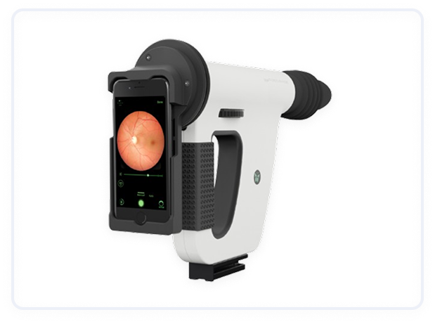
Fundus Cameras
A fundus camera is a specialized ophthalmic imaging device used to capture high-resolution images of the fundus, or the interior surface of the eye, including the retina, optic disc, macula, and blood vessels. It plays a crucial role in diagnosing, monitoring, and managing a variety of eye diseases and systemic conditions that affect the retina and optic nerve.
Benefits of Fundus Cameras
Non-invasive:
Fundus cameras offer a non-invasive way to capture detailed images of the retina, making the procedure comfortable for patients without the need for surgery or incisions.
High-Resolution Imaging:
Fundus cameras provide high-resolution images, allowing clinicians to detect even small changes in the retina, which can be crucial for early diagnosis and treatment of various eye conditions.
Wide Field of View:
Modern fundus cameras can capture a wide field of view of the retina, providing a comprehensive assessment of the central and peripheral retina in a single image.
Early Detection and Monitoring:
The ability to track changes over time makes fundus cameras an essential tool for monitoring the progression of diseases like diabetic retinopathy, glaucoma, and macular degeneration.
Easy Documentation:
The digital nature of fundus imaging allows for easy storage, sharing, and comparison of images across time, enabling better management of patient history and treatment plans.
Automated Analysis:
Some modern fundus cameras have integrated software that can automatically analyze images and detect signs of disease, offering a faster, more efficient way to assess the retina.
Types of Fundus Cameras
Non-Mydriatic Fundus Cameras:
These cameras allow for imaging of the retina without dilating the pupil. They are easier to use in a clinical setting because they eliminate the need for eye drops. Non-mydriatic cameras use specialized wide-angle lenses to capture a broad field of view of the retina.
Mydriatic Fundus Cameras:
These cameras require dilated pupils to provide a wider field of view and sharper images. Mydriatic cameras are typically used in detailed examinations or when a more comprehensive view of the retina is necessary.
Portable Fundus Cameras:
These are compact, handheld devices, often used in field settings or for quick screening. They are particularly useful for mobile healthcare units or for remote regions where access to advanced ophthalmic equipment might be limited.
Types of Fundus Cameras
Non-Mydriatic Fundus Cameras:
These cameras allow for imaging of the retina without dilating the pupil. They are easier to use in a clinical setting because they eliminate the need for eye drops. Non-mydriatic cameras use specialized wide-angle lenses to capture a broad field of view of the retina.
Mydriatic Fundus Cameras:
These cameras require dilated pupils to provide a wider field of view and sharper images. Mydriatic cameras are typically used in detailed examinations or when a more comprehensive view of the retina is necessary.
Portable Fundus Cameras:
These are compact, handheld devices, often used in field settings or for quick screening. They are particularly useful for mobile healthcare units or for remote regions where access to advanced ophthalmic equipment might be limited.
Various industries
Request More Information
Contact Us to learn more about our offerings or Request a Quote to get started today!
Contact Us

